
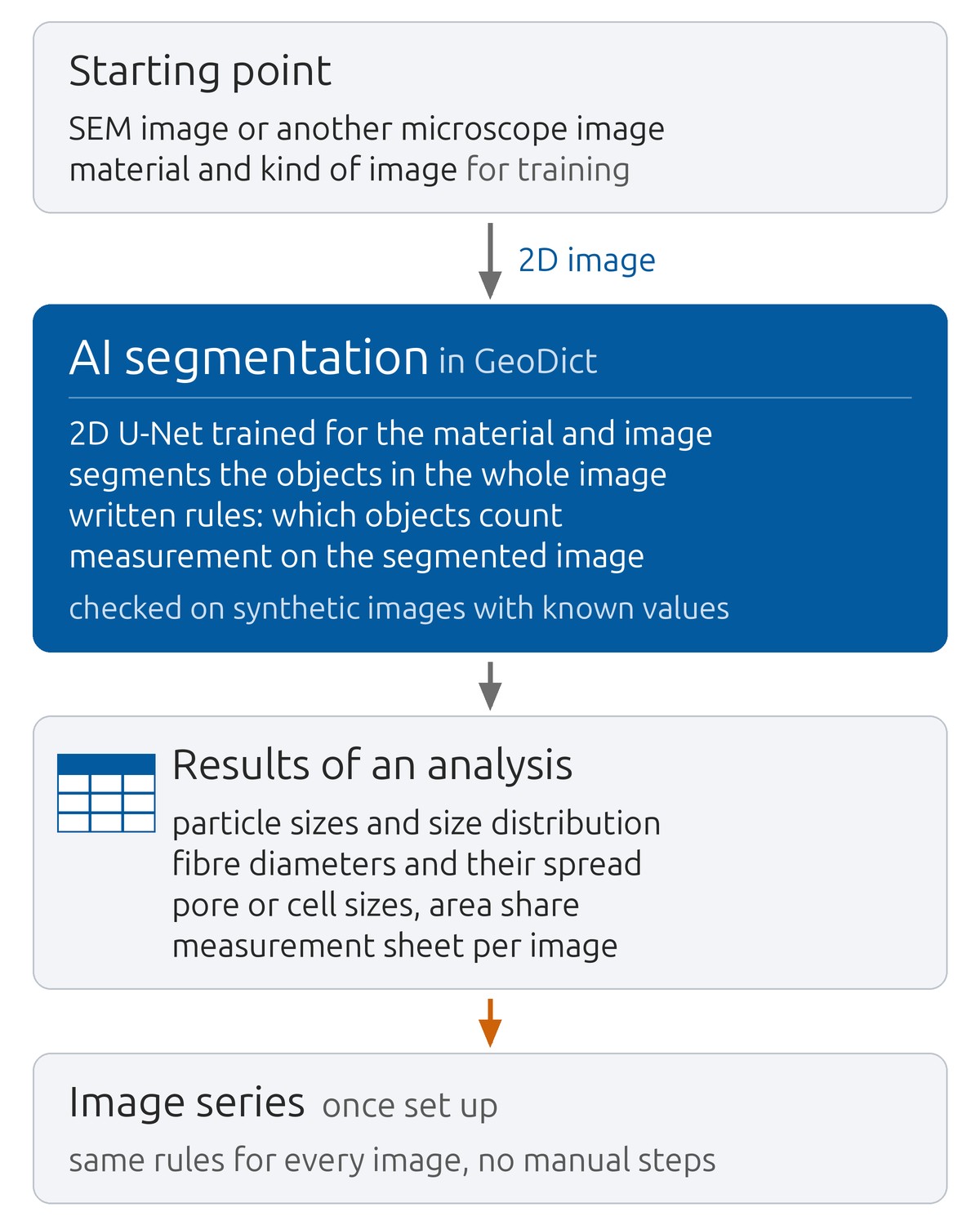
Measuring the sizes of particles, fibres or pores in a microscope image by hand takes time, and the result depends on who measures. With a grey value threshold, objects are separated from the background only where they differ in brightness. In GeoDict, the software for digital material R&D by Math2Market, a neural network for image segmentation can be trained for a material and a kind of image, and the objects in the whole image are segmented with it.
Results of an analysis
- Particle sizes, shapes and size distribution.
- Fibre diameters and their spread.
- Pore or cell sizes and their area share.
- A measurement sheet per image with the measured objects marked, the values, and each value’s place in the image.
AI segmentation and checks
The model is a 2D U-Net, and each object is measured on the segmented image. Before use, the measuring chain is checked on synthetic images with known true values. A size measured in a section or projection is not the 3D size of the object, and pore connectivity cannot be read from one image.
Image series
Once set up, the measuring chain runs per image without manual steps and suits series of many images, for example in an analysis laboratory. Which objects count and which are rejected, and why, is written down and applied in the same way to every image.
Examples
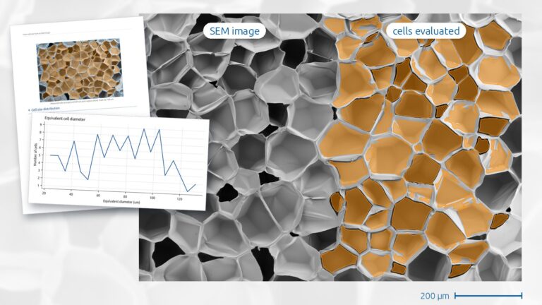
In the case study, the cells of a foam are measured in one SEM image that was among the training images of the model, so the result is no test on an unseen image.
Analysis of microscope images
cvt engineering offers the analysis of SEM images and other 2D micrographs as a service. The segmentation model is trained on images of your material of the kind to be measured.
Related pages: AI-assisted development of filter media and nonwovens, Digital development of porous transport layers (PTL) for PEM electrolysis, Digital twins of ceramic membrane coatings from nano-CT