
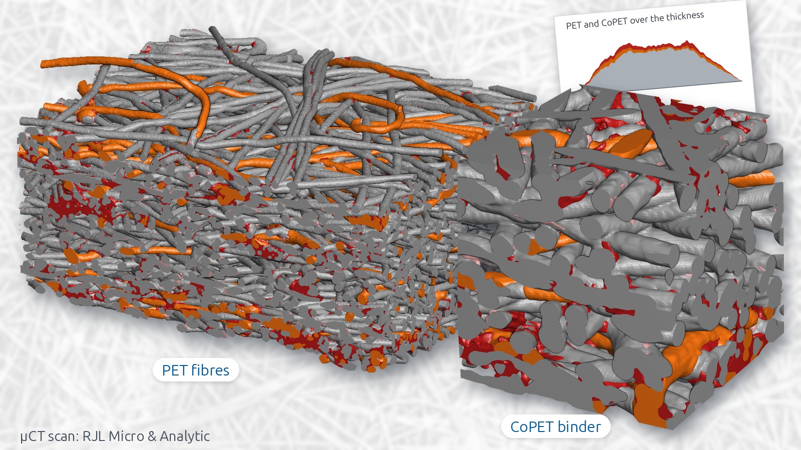
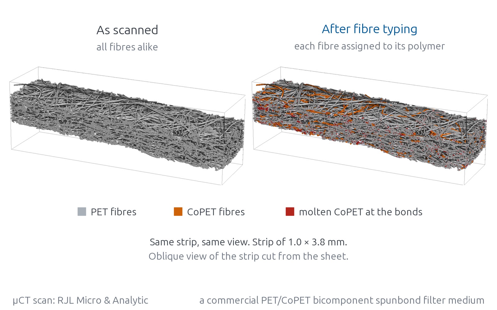
This spunbond is made of two polyesters: PET fibres that carry the sheet and CoPET fibres that melt in the calender and bond it. In a CT scan the two look alike, so a segmentation by grey value sees one material. Here they are told apart in a µCT scan, which gives how much binder the sheet holds and where it sits.
The sample
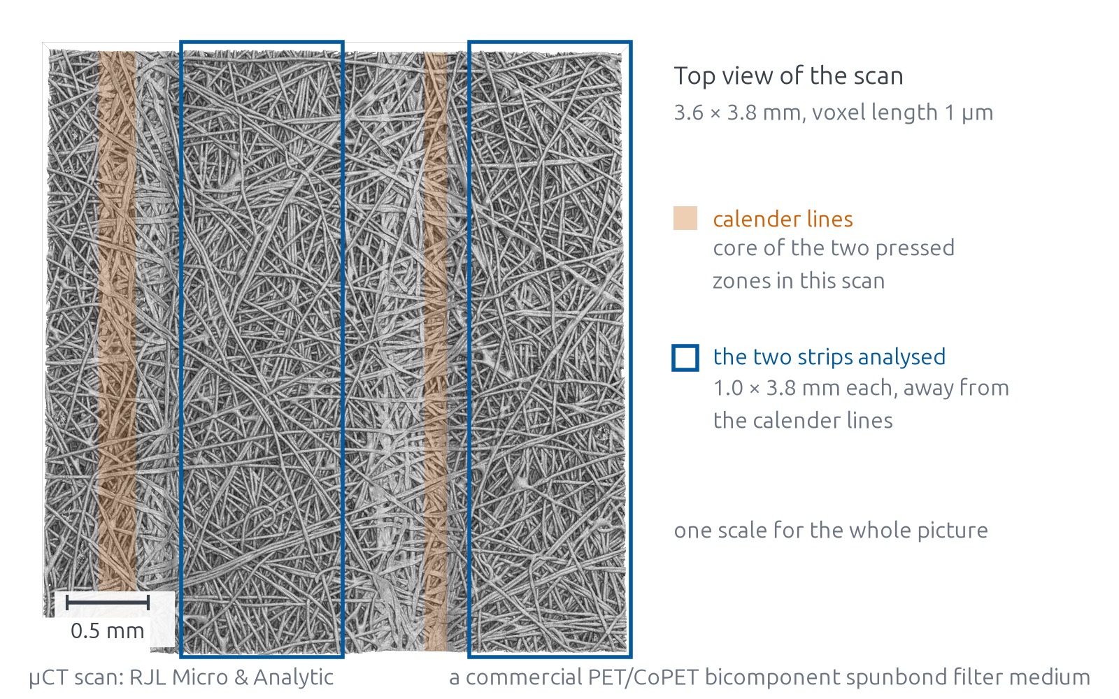
The sample is a commercial PET/CoPET bicomponent spunbond filter medium, thermally bonded in a calender. RJL Micro & Analytic scanned a piece of a few millimetres in a µCT. Two strips away from the calender lines were analysed. The case study on preparing a CT scan uses a cut-out of the same scan at a coarser resolution.
Binder fibres and bonds
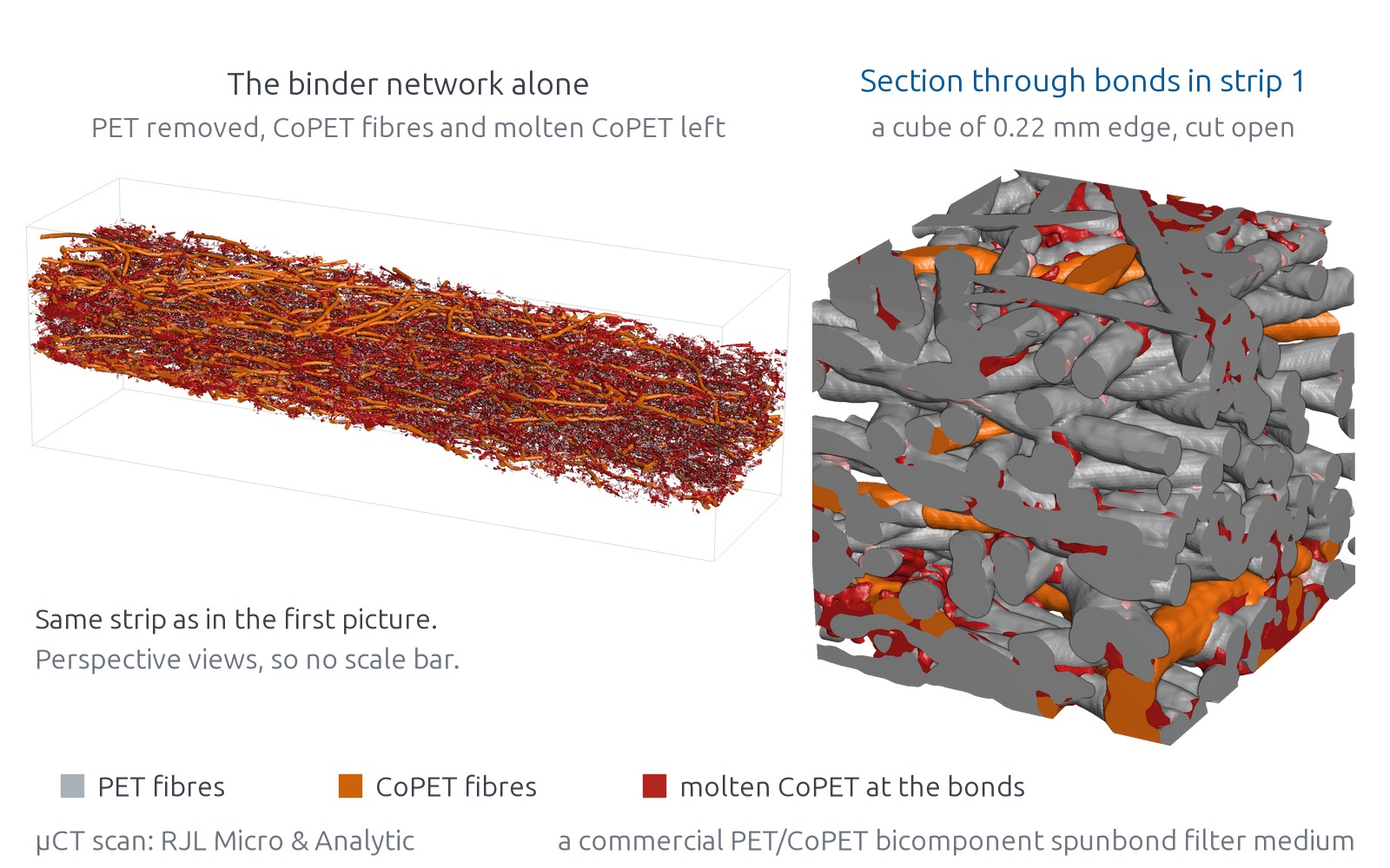
With the PET removed, the binder stands alone: CoPET fibres that kept their shape, and CoPET that melted and now sits at the contacts between fibres. Every analysed PET fibre in the two strips is bonded.
Binder share
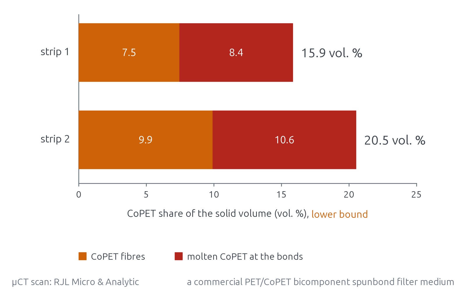
The CoPET share is given as a share of the solid volume, for the fibres and the molten material separately. It is a lower bound, because CoPET that has melted into films thinner than the scan resolves is not counted. The two strips lie on either side of a calender line and give different shares; part of the difference lies within the uncertainty of the evaluation.
Binder across the thickness
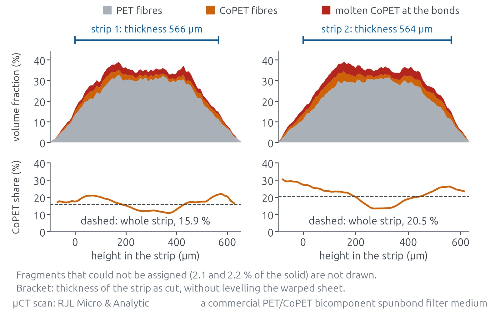
Across the thickness the CoPET share is higher towards both faces of the sheet than in its middle.
The structure of the sheet
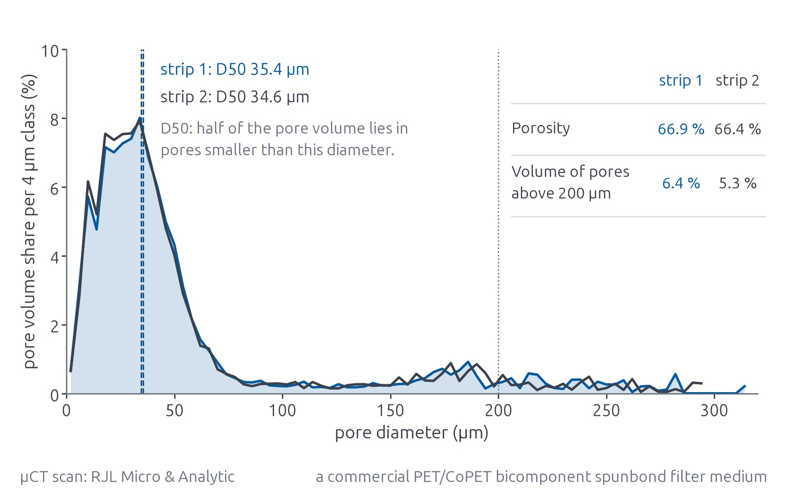
The same strips give pore sizes and porosity.
Limits
The scan covers a few square millimetres of one sheet and does not describe a roll. The CoPET share is a lower bound. The method has been applied to this one material so far. The assignment of the fibres has not been checked against a chemical analysis of single fibres. Filtration has not been simulated on this structure.
Fibre analysis of nonwovens
cvt engineering offers this analysis for CT scans of other thermally bonded nonwovens with carrier and binder fibres.